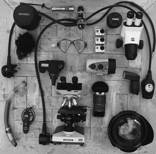
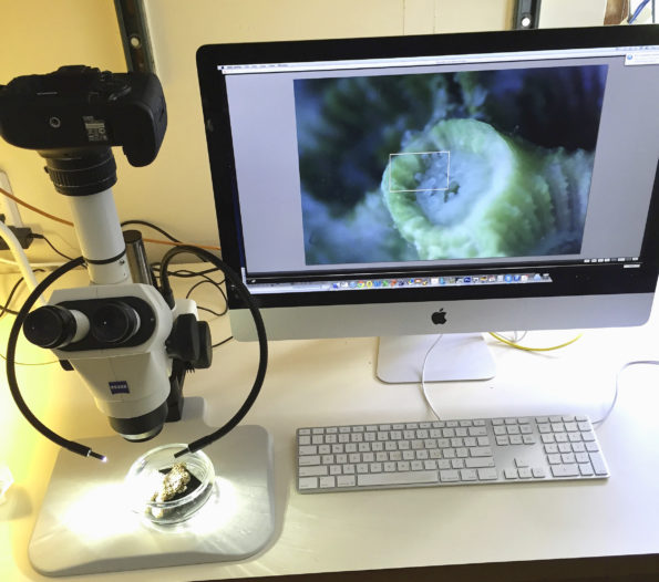
The image below (left) shows one of two microscopes that we use on our expeditions. The camera is a Canon 5D Mark III and can interface with the computer. In live view mode the computer displays what the camera “sees” in realtime. The camera can be controlled from the keyboard. This stereo microscopes acts like a high-powered zoom lens with a range of 1:1 to 7:1 and allows the photographer to fill the screen with a single coral polyp. The coral on the computer screen (and in the finger bowl) is Astrangia poculata commonly known as the Northern Star Coral. The specimen was collected by the Marine Resources Department of the Marine Biological Laboratory in Woods Hole, MA. The image was taken by Joe in his laboratory at the MBL in the summer of 2012. The image on the right shows some of our gear that we bring with us. This includes two types of microscopes both compound and stereo microscopes, GoPros, underwater housing for Canon 5D Mark IV with strobe and video lighting.

